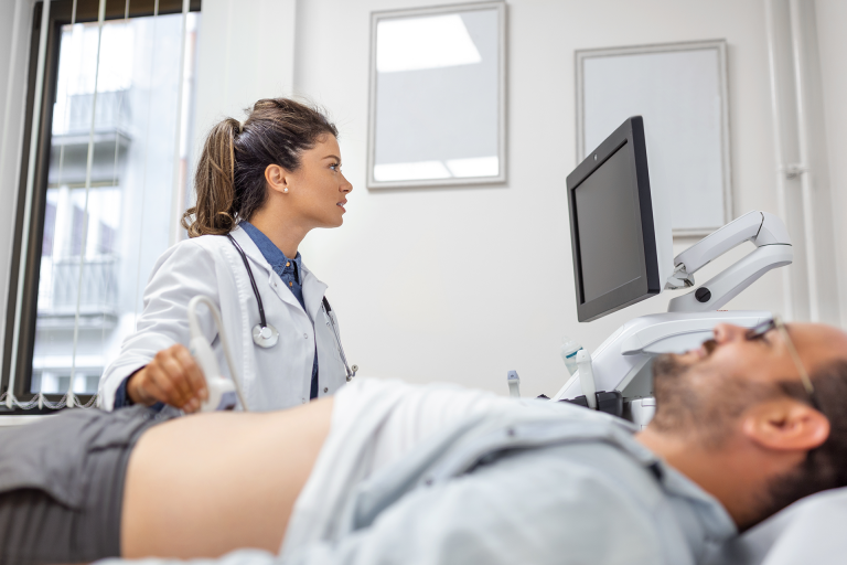
Testicular ultrasound at “Republikas laukuma klīnika” in the center of Riga is a highly precise, safe, and entirely painless diagnostic procedure used to evaluate male reproductive health. This non-invasive examination uses advanced sound wave technology to provide detailed, real-time images of the testicles, epididymis, and surrounding tissues. Whether used for a routine health check, investigating localized pain, or as part of a fertility assessment, our specialists ensure a professional and comfortable environment.
Testicular ultrasound | Republikas laukuma klīnika
Testicular ultrasound is a painless imaging method that provides important information about male reproductive health. This examination allows assessment of the morphology and functional condition of the testicles, their appendages, and the initial part of the spermatic ducts.
Ultrasound is especially important for men with fertility issues, as it helps diagnose various problems related to testicular function.
At “Republikas laukuma klīnikā”, testicular ultrasounds are performed by highly qualified specialists who focus on reproductive health, ensuring accurate diagnostics and, if necessary, further treatment.

Dr. Vladimirs Šalajevs
Principle of testicular ultrasound
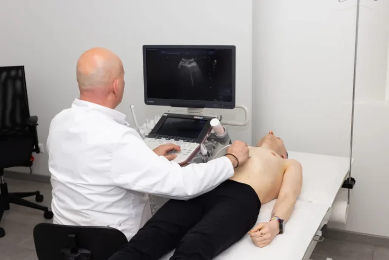
Testicular ultrasound (USG) is a non-invasive medical procedure that uses sound waves to create images of the testicular tissue. During the procedure, a special gel is applied to the skin to ensure good contact between the ultrasound probe and the skin.
The probe emits sound waves into the tissue and detects the reflected signals. These signals are converted into digital images that show the internal structure of the testicles. Unlike X-rays, ultrasound does not use ionizing radiation, making it safe and repeatable as needed.
Modern USG devices provide high-resolution images, allowing detailed evaluation of testicular condition. Doppler technology can also assess blood flow within the testicular tissue.
Testicular anatomy and importance of ultrasound
The testicles are paired organs located in the scrotum. They produce sperm and male hormones.
Testicular ultrasound allows assessment of their size, shape, and internal structure. It helps diagnose varicocele (vein enlargement), hydrocele (fluid accumulation), testicular tumors, and inflammation.
Ultrasound is especially important in fertility investigations, as it can detect reduced sperm count, motility, and abnormal forms. The procedure is painless and lasts about 15–20 minutes, providing quick and accurate information about testicular health.
Testicular USG procedure
No special preparation is usually required. Some recommendations to improve quality:
- Wear comfortable, easily removable underwear and loose clothing.
- Inform the doctor about existing testicular problems or previous injuries.
- Provide information about any medications you take regularly.
A full bladder is not necessary. The doctor will explain the procedure and answer questions beforehand.
Procedure steps
- Preparation: The doctor applies a special gel to the scrotal skin for better sound wave conduction.
- Scanning: The ultrasound probe examines both testicles, appendages, and surrounding tissue.
- Documentation: Images are taken from multiple angles to evaluate size and structure.
Patients may feel slight pressure, but discomfort is minimal.
Interpretation and possible findings
Testicular USG can detect:
- Masses: Cysts, tumors (benign or malignant)
- Structural changes: Size changes, calcifications, or scarring
- Circulation issues: Varicoceles, inflammatory processes
The doctor evaluates testicular size, structure, blood flow, symmetry, as well as the epididymis and scrotum. Results are usually available immediately, and further action—follow-up, additional tests, or treatment—will be recommended if necessary.
Frequently asked questions
When is testicular ultrasound needed?
- Pain or discomfort in the testicular area
- Swelling, hardness, or palpable lumps
- Asymmetry or sudden changes in size or shape
- Red, warm skin or chronic groin pain
- Part of infertility investigations or after groin/testicular injuries
How to prepare for testicular ultrasound?
- No special preparation is needed.
- Expose the lower abdomen and groin area during the procedure.
- Wear comfortable, easily removable clothing.
Cost of testicular ultrasound
The cost of testicular ultrasound at “Republikas laukuma klīnikā” izmeklējuma cena ir €70,00.