Neurosonography in infants | Republikas laukuma klīnika
Neurosonography in infants (also called neurosonological ultrasound or neurography in infants) is one of the most important and safest methods used by pediatricians and neonatologists to assess the brain development of newborns and infants. It is an ultrasound examination method that allows detailed visualization of brain structures through the fontanelle, while it is still open. This examination is painless, harmless, and provides essential information about a child’s health.

Dr. Jeļena Liepa
What is neurosonography in newborns?
Neurosonography is an ultrasound method that allows evaluation of an infant’s brain structures. It is performed through the fontanelle – a natural opening in the skull that remains unclosed during the first months of life. The examination helps to detect possible developmental abnormalities, hemorrhages, fluid accumulation, or other pathologies at an early stage.
Unlike other imaging methods, such as computed tomography or magnetic resonance imaging, neurosonography in infants is completely harmless and suitable even for premature babies. It can be repeated several times to monitor the child’s development.
Why is neurosonography necessary?
Neurography in infants is essential because it helps the pediatrician and neonatologist to diagnose and prevent serious health problems in time. The main reasons for prescribing neurosonography are:
- for premature babies, to assess brain development;
- if complications occurred during childbirth (oxygen deprivation, trauma);
- if the child has an unusual head shape or size;
- if developmental delay or neurological symptoms are observed;
- if congenital brain anomalies are suspected;
- as a preventive measure, to confirm normal brain development.
How is neurosonography performed?
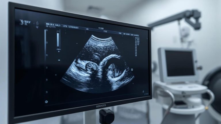
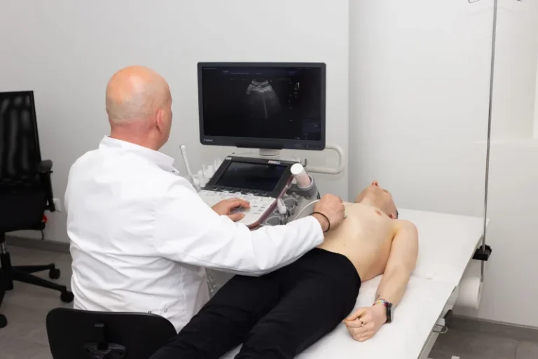
The examination is simple and painless. During the procedure, the baby is placed on their back, and the doctor uses an ultrasound probe to examine the brain structures through the fontanelle. The examination lasts 10–20 minutes, and the baby usually remains calm, as no anesthesia or special preparation is required.
Neurosonological ultrasound allows the doctor to see the ventricular system of the brain, blood vessels, white and gray matter structures, as well as possible lesions or fluid accumulation.
Preparation for the examination
No special preparation is required. However, it is recommended that the baby is fed and calm, as this makes the examination easier. Parents should bring the child’s medical records so that the pediatrician or neonatologist can evaluate the results in the context of the child’s health history.
What does neurosonography reveal?
Neurosonography helps diagnose various conditions:
- intraventricular hemorrhages,
- hydrocephalus (fluid accumulation in the brain),
- brain structural anomalies,
- ischemic lesions,
- infection‑related changes,
- tumors or cysts.
Early diagnosis allows treatment to begin and prevents complications that could affect the child’s long‑term development.
The role of the pediatrician and neonatologist
The pediatrician is the first specialist parents turn to with questions about their child’s health. They assess the child’s development and, if necessary, refer them for neurosonography.
The neonatologist is a doctor specializing in newborn care. They often prescribe neurosonography for premature babies or newborns with complicated birth histories. Both specialists work closely together to ensure the best possible diagnosis and treatment for the child.
Advantages of neurosonography
- Safe and harmless method.
- Painless and quick procedure.
- Widely available in medical institutions.
- Provides precise information about brain development.
- Can be repeated multiple times to monitor changes.
When should the examination be repeated?
In some cases, neurosonography needs to be repeated to evaluate brain development over time. For example, in premature babies, the examination is performed several times during the first months of life. The pediatrician or neonatologist determines the frequency of repetition depending on the child’s health condition.
Safety and parents’ concerns
Parents often have questions about the safety of the examination. Neurosonography is completely harmless because it uses ultrasound waves, not radiation. It can even be performed several times a month if necessary. The baby does not feel pain during the procedure, and there is no risk of complications.
Neurosonography in infants is a safe and effective diagnostic method used by pediatricians to assess brain development. This examination is also called neurosonological ultrasound or neurography in infants. It helps to detect developmental disorders, hemorrhages, or fluid accumulation in the brain at an early stage. The procedure is painless, harmless, and suitable even for premature babies. Parents do not need to worry about preparation – it is enough for the baby to be fed and calm. The results are evaluated by a pediatrician or neonatologist to ensure the best possible care for the child.
Frequently Asked Questions
Is neurosonography safe?
Yes, it is a completely harmless and painless method.
How long does the examination take?
Usually 10–20 minutes.
Is preparation required?
No special preparation is needed, but the baby should be fed and calm.
Who evaluates the results?
The results are analyzed by a pediatrician or neonatologist.
Can the examination be repeated?
Yes, it can be performed multiple times if it is necessary to monitor the child’s development.